How Does Skin Cancer Screening Work
Skin Cancer Screening Saves Lives
Getting a Skin Cancer Check with “Mole Map”
“What are your plans for the day?” the tall brunette asked, as she rung up my Lulu Lemon shopping spree (I needed some new running shorts! They didn’t disappoint).
“I’m actually headed around the corner for a ‘mole mapping’, have you heard of that technology?”, I offered, excited to hear her reply.

Anxiously, she replied “Yes, you must be nervous! I’d be so scared of what they might find!”
I chuckled, “Well, actually I’m a bit used to it by now”, pointing to my Health Nucleus jacket and explaining this wasn’t my first cancer screening experience, quite the opposite in fact!
“Well, I’m sure you’ll be fine”, she offered.
“Actually, that’s what the appointment is for”, my somewhat cheeky reply.
“Don’t worry, be happy”
In her polite laugh, I could see the point had landed.
“The clinic is just around the corner, you should check it out sometime”, my closing remark, as I headed off to a comprehensive skin cancer screening service, in search of that ‘sure you’ll be fine’ level of certainty - but not on a whim, instead, at the guiding hand of longevity technology.
Skin cancer spreads and kills

Bob Marley - legendary musician, skin cancer victim
When Bob Marley, one of the most influential musicians of his time, collapsed during a run around Central Park, his first thought was not likely to fault skin cancer for the scare.
But further analysis revealed that a known cancerous growth in the skin under his big toenail had metastasised, spreading to his brain, liver and lung. He was dead only a few months later at age 36.
His death shook the world in a way not unlike that of Chadwick Bosemen, who died a similarly tragic death at the age of 43 from colorectal cancer last year, raising awareness of the disease, which has unfortunately continued to reek havoc on the lives of millions of people each year.
In my home country of Australia, skin cancer is particularly rife. 2 out of every 3 Australians will get some form of skin cancer by age 70. On average, an Australian dies every 5 hours from melanoma, the most aggressive and dangerous form of skin cancer.
However, it is not just older folks who are affected. Skin cancer is one of the most common cancer diagnoses for younger people in the ages of 25-44.
And while the 1970s did not offer the types of advanced skin cancer screening technologies that could have saved Bob Marley’s life - 2021 proves much different.
You don’t have to leave your skin cancer risk up to chance. You can manage the risk.
Let’s look at how.
 |
Skin cancer screening saves lives
As I explain in the above video, my visit to a MoleMap clinic in the Sydney CBD was to provide a comprehensive skin cancer screening, leveraging imaging technology to make a ‘map’ of my skin, forming a baseline for future tracking.
The MoleMap clinic utilised a few key pieces of technology to complete this screening.
An example of one of the 26 high resolution images taken of my body.
Step 1: Full Body Images
The first was a series of high resolution photos, where, stripped ‘down to me undies’ (said in the lovely Irish accent of my melanographer Kathy).
In a series of coordinated movements, using a reference chart on the floor for my foot positions, I posed for more than 25 images.
These were taken with a Nikon DSLR, set for maximum focus on the skin of the body.
Each image was then mapped onto a virtual avatar in a specialised computer system.
Unfortunately the avatar was much more ripped than I, perhaps I will equal his masculinity next visit?
Step 2: Head to Toe Examination
After the full body images were taken, the melanographer (who was a registered nurse, specially trained by the Australiasian College of Dermatologists) reviewed my body from head to toe.
Side Note: I wasn’t asked to take off the underwear, unless I had a mole or skin issue I wanted them to track. Ladies - bra is optional, but recommend that you go without! Your female melanographer will be sure to make you comfortable and there is a robe you can use to cover up.

The dermatoscope in action. This tool is used after a mole has been marked by visual inspection, to further screen its shape, colour and size.
In this head to to review, Kathy would look at each of my moles using a Dermatoscope
This handy gadget uses a LED lighting and a magnifying lens to assess the surface features of a mole (more broadly referred to as ‘lesions’).
It allows up to 2.5x magnification and allows for the colour, shape and size to be reviewed in more detail.
Any more that ticks the boxes of being at increased risk (more on that in a moment), would have its location ‘tagged’ on my virtual avatar.
Kathy reviewed me thoroughly, including within my hairline, in a highly professional and considerate way.
Step 3: Enter the MoleCam
For any mole displaying any of the characteristics of skin cancer (basal cell carcinoma, squamous cell carcinoma or melanoma), further imagery was take using specialised device aptly named the “MoleCam”.
The dermatoscope identifies a mole with asymmetry and irregular border on my leg.
To be clear - this is not to say these moles are likely to be cancerous.
Instead, they match known characteristics of skin cancer risk are best remembered by the “ABCDs”:
Asymmetry (irregular shape)
Border (ragged or uneven)
Colour (more than two shades)
Diameter (greater than 6mm)
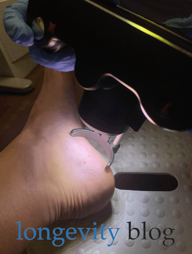
For each at risk mole, the MoleCam was used to capture two images.
The first, at a set distance and focal length - you can see the ‘slide’ extended from the device.
Second, with a ‘contact’ dermascopic image, where a dab of alcohol is placed on the lens, and the MoleCam is placed directly against the skin.
In the below 3x images, from left to right, we can observe the process.
The first image at extended focal length. The second ‘contact’ image. And finally, the contact image of the mole on my left foot is displayed on the device - you can see that Kathy rightly spotted its irregular shape, border and colour.


Step 4: Teledermatologist Review
In total, I had 26 moles of interest tagged and submitted for further review.
This is above average, and does suggest that my longevity is at elevated risk for skin cancer.
In general the moles were present all over my body, including the back, chest, abdomen, legs, arms and feet.
In some areas, sun exposure did appear to play a role, but in others, my naturally ‘mole-y’ complexion likely drove their prevalence.
Each of the detailed images that were taken by the MoleCam will now head off for a review by a ‘teledermatologist’, which is pretty much a fancy way of saying that a skin cancer expert will review each of the moles.
After this review, there will effectively be a ‘ranking’ of the risk each of the moles represents.
I will then receive customised advice on the MoleMap online patient platform, about 7 days or so after the appointment.
In most cases, the actionable information will be - watch & wait.
This is because the 5th aspect of the ABCDs of skin cancer is actually an E, standing for Evolution, or changes over time.
Cancerous moles, due to unregulated growth driven by DNA damage, generally have an inability to heal and fast cell turnover.
Whereas normal skin/moles will be steady-state, skin cancers will be changing. This is why your first MoleMap visit is so important - you form a baseline by which to assess the Evolution of your unique mole makeup.
This brings us to Step 5!
Step 5: Monitoring for Skin Cancer
The brilliant aspect of this mole mapping longevity technology is the ability to track changes in your moles over time.
New moles, quick changing moles or any areas of the skin which begin to react to excessive sun damage, need to be re-assessed over time.
Your MoleMap appointment will provide you with valuable reference material for self-assessment and tracking at home.
Depending on your individual risk factors (age, sun exposure, existing moles, genetics), MoleMap will provide you with guidance on how often you should re-assess with a follow-up appointment.
By the way - you can check your personal risk with this handy tool on the MoleMap website.
This of course also depends on your budget, and is one reason why you should form a personalised longevity strategy, and include skin cancer screening as a part of your approach to life a long and healthy life.
For me, this will be an annual visit (every 12 months), supplemented by a self-exam every 3 months.
Since I now know where each of my moles on interest are, and have been provided with some handy tools by the MoleMap team.
This includes a thorough information sheet, which I’ve uploaded here (see image).
However, one interesting fact to keep in mind - you can only ‘see’ about 40% of your skin - so be prepared to ask for some help!
Skin Cancer Screening - the Future
Coming up in our next post, A Longer Life will interview MoleMap’s Chief Customer Officer Vlad Mehakovic.
We’ll dive deeper into how you can use skin cancer screening technology to manage your longevity risk, as well as chat about where this longevity technology will go in the future!
Be sure to subscribe below, so you won’t miss it!


FDA & TGA DISCLAIMER
This information is intended for educational purposes only and is not meant to substitute for medical care or to prescribe treatment for any specific health condition. These blog posts are not intended to diagnose, treat, cure or prevent any disease, and only may become actionable through consultation with a medical professional.